|
Although they are excreted far from the brain, many EVs from cancer cells exist stably and are excreted in the urine without breaking down. Urine testing has many advantages, explains Associate Professor Takao Yasui of Nagoya University Graduate School of Engineering. “Liquid biopsy can be performed using many body fluids, but blood tests are invasive,” he said. “Urine tests are an effective, simple, and non-invasive method because the urine contains many informative biomolecules that can be traced back to identify the disease.”
|
|
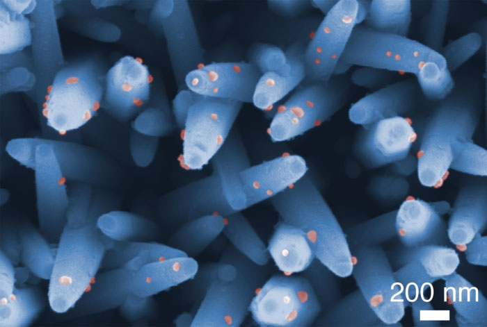
A research group led by Yasui and Professor Yoshinobu Baba of Nagoya University’s Graduate School of Engineering, in collaboration with Nagoya University’s Institute of Innovation for Future Society and the University of Tokyo, has developed a new analysis platform for brain tumor EVs using nanowires at the bottom of a well plate. Using this device, they identified two specific types of EV membrane proteins, known as CD31/CD63, from urine samples of brain tumor patients. Looking for these tell-tale proteins could enable doctors to identify tumor patients before they develop symptoms.
|
|
“Currently, EV isolation and detection methods require more than two instruments and an assay to isolate and then detect EVs,” said Yasui. “The all-in-one nanowire assay can isolate and detect EVs using one simple procedure. In the future, users can run samples through our assay and change the detection part, by selectively modifying it to detect specific membrane proteins or miRNAs inside EVs to detect other types of cancer. Using this platform, we expect to advance the analysis of the expression levels of specific membrane proteins in patients’ urinary EVs, which will enable the early detection of different types of cancer.”
|